Thursday, October 4, 2018

SCIREQ Blog has moved!


Thank you for following the SCIREQ Blog. 


We moved our Blog to a new portal and invite you to continue reading our posts and stay informed about:

         »        Events & User Group Meetings
         »        Products Developments
         »        Recent Publications
         »        and news from the pulmonary community.


Follow this link to visit SCIREQ Blog!

Happy Reading!

Your SCIREQ Team 

Monday, July 23, 2018

FASEB’s Lung Epithelium in Health and Diseases conference


SCIREQ is proud to sponsor FASEB’s Lung Epithelium in Health and Diseases conference which will take place from July 29th to August 3rd, 2018 in St. Bonaventure, NY. The scientific research conference will bring together leading scientists and junior researchers to discuss the latest findings in the field of lung epithelium biology. 

The conference program touches on various respiratory ailments, disease models and novel research techniques, including:

  • Mucociliary transport in cystic fibrosis (Michael Welsh, University of Iowa)
  • Lung development and regeneration (Jeffrey Whitsett, Cincinnati Children’s; Bernard Thébaud, University of Ottawa)
  • Neuroendocrine Hyperplasia of Infancy (Lisa Young, Vanderbilt University)
  • Disease modelling with organoid culture systems and cryopreserved human lung slices  (Xingbin Ai, Brigham & Women’s Hospital; Barry Stripp, Cedars-Sinai)
  • Genetics and Genomics of Lung Diseases (Steve Rowe , U.of Alabama; David Schwartz, U.of Colorado)
  • And many other topics

Links:
Program: https://secure.faseb.org/src-programs/11508.pdf  
Conference website: http://faseb.org/src/micro/Site/Lung/Home.aspx 

We look forward to talks, poster sessions and new collaboration opportunities that will emerge from the FASEB event.

Thursday, July 5, 2018

Bronchopulmonary Dysplasia (BPD) after 50 years – BPD Symposium at Stanford

A momentous symposium was held at Stanford university last year to commemorate the 50th anniversary since the original description of Bronchopulmonary Dysplasia (BPD) was first articulated1 by Dr. William Northway and him team in 1967. 

This symposium, which was attended by several leaders in the field, was held as a platform where researchers could come together and highlight the progress that has been made to date, along with discuss future perspectives. 

The event was divided into 5 sessions with keynote presentations provided by speakers who have made noteworthy contributions in the field of BPD. We are proud to say that amongst such speakers were Dr. Bernard Thébaud, Dr. Rory Morty and Dr. Rob Tepper, who gave very informed and thought provoking lectures during the symposium, are active users of our SCIREQ systems!

We would like to extend our Thank You to all investigators who constantly strive towards making new strides in this field of preclinical pediatric research.

Learn more about how SCIREQ’s solutions can be implemented into your research needs or how our instruments are being utilized to study Bronchopulmonary Dysplasia


1Northway et. al. Pulmonary Disease Following Respirator Therapy of Hyaline-Membrane Disease — Bronchopulmonary Dysplasia, N Engl J Med 1967; 276:357-368


Friday, June 22, 2018

IDEAL OR ACTUAL BODY WEIGHT OF THE OBESE SUBJECT?

When working with obese mice, what weight should be used when performing flexiVent measurements?


This question is quite an important one for two reasons. First, there is a substantial difference in weight between the obese subjects vs. the lean control group. For instance, obese mice can weigh 50g or more, while their age-matched control group can typically weigh 30g. While there is this significant difference in the weight between both groups, there may not be a large difference in the actual size of the subjects’ lungs.

Second, when working with the flexiVent system, the body weight of the subject is used to scale the amplitude of volume-controlled ventilation patterns and perturbations, such as the Snapshot-150 or the Quick Prime-3. In obese animals, the increase in body weight is not proportional to the subject’s lung size, so sending in a larger volume can have adverse effects on the subject’s lungs.


A recent study performed by Dr. Guivarchis et al.1, highlights the harmful effects of ventilating obese subjects with a tidal volume based on actual weight vs ideal weight using the flexiVent system. In this study, the effects of two hours of mechanical ventilation in a diet-induced obese mice model, with tidal volume calculated on either the actual body weight or the ideal body weight (based on the mean weight of control mice) were demonstrated. Their findings indicate that in comparison to lean control subjects, mechanically ventilating obese subjects with a tidal volume based on actual body weight is harmful.

They observed a noticeable variation in lung mechanics and associated lung inflammation in obese mice ventilated with a tidal volume based on actual body weight. In contrast, obese mice ventilated with a tidal volume calculated using the ideal body weight had lung mechanics and inflammation parameters close to the lean control group.

Therefore, when studies involve the assessment of obese mice, SCIREQ recommends entering the average weight of the lean control group rather than using the actual weight of the obese subjects.


1Guivarch et al. Pulmonary Effects of Adjusting Tidal Volume to Actual or Ideal Body Weight in Ventilated Obese Mice. Sci Rep. 2018 Apr 24;8(1):6439. doi: 10.1038/s41598-018-24615-5.

Monday, June 11, 2018

To open or close the lungs – that is the question!

Although often necessary, mechanical ventilation of small animals during pulmonary measurements can lead to both acute and chronic damage to their lungs, also known as ventilator-induced lung injury (VILI). The injury happens as a result of the frequent opening and closing of alveolar air sacs. Nevertheless, the two most common, and greatly contrasting methods, to study respiratory diseases include open lung- and closed lung-approaches. This has led to many pulmonary researchers conducting experiments with the goal of determining which approach is the least harmful and the best possible way to minimize VILI.

TO OPEN THE LUNGS?

Open-chest models are often used to study ARDS and acute myocardial ischemia and reperfusion. These models allow for investigating cardiac physiology, morphological changes of the lungs, as well as the evaluation of therapeutic interventions. This procedure allows for invasive lung function measurements and tissue harvesting for further experimentation, but must be performed under deep anesthesia. Since opening the chest causes the lungs to collapse, the animals must be mechanically ventilated throughout the process.

The flexiVent has often been used in open-chest experiments, as the system allows for ventilation of the subject with a user-defined positive end-expiratory pressure (PEEP), which can be set to prevent airway collapse. Further, it can facilitate the complete recruitment of the lungs, using the Deep Inflation perturbation, before measurements can be taken.


TO CLOSE THE LUNGS?

Recent experimental evidence suggests that high PEEP does not necessarily reduce lung injury but instead, may lead to the contrary. Higher PEEP results in overstretching the alveolar sacs and potential development of edema, among other cardiopulmonary impairment. Moreover, a recent article by Dr. Patricia Rocco and team1, points out that most publications comparing high PEEP with no PEEP, do so with a combination of low tidal volume and high tidal volume, respectively. Thus, there is no clear indication as to what exactly led to the observed outcomes – high PEEP or low tidal volume.

Dr. Rocco goes on to recommend “in order to minimize VILI, we should consider moving away from the classical concept of ‘open up the lungs and keep them open’ towards ‘close down the lungs and keep them closed’”. Fortunately, the flexiVent can be adapted to different experimental conditions, and can provide detailed pulmonary measurements even as research trends change.


1Pelosi, P. et al. 2018. Close down the lungs and keep them resting to minimize ventilator-induced lung injury. Critical Care 22 (1):72.

Monday, May 14, 2018

Must see posters at ATS!

ATS is just around the corner and we hope to see you in San Diego!

We are looking forward to learning more about respiratory research, especially these interesting flexiVent poster sessions:

During our annual breakfast event at ATS on Monday, May 21st, Dr. Otmar Schmid (Helmholtz Zentrum, Germany) will discuss his “off-label” use of the flexiVent for the delivery of inhaled compounds. By optimizing the ventilation profiles and nebulizer settings, Dr. Schmid and Dr. Annette Robichaud obtained significant improvements in aerosol deposition rates, as well as homogenous deposition profiles.

The technique developed by Dr.Schmid could be useful in studies for optimized inhaled drug delivery using the flexiVent. If you are interested, please feel free to join us by registering here.

We would love to learn more about your research and discuss some tailored solutions. Find us at Booth #1535 in Exhibit Hall D.

Exhibit Hours

Sunday, May 20       10:30 am – 3:30 pm
Monday, May 21      10:30 am – 3:30 pm
Tuesday, May 22      10:30 am – 3:30 pm


Monday, April 16, 2018

Cardiopulmonary Solutions by emka & SCIREQ

emka & SCIREQ offer a unique perspective into the preclinical study of heart and lung diseases, allowing for novel insights into cardiopulmonary diseases such as heart failure, arrhythmia, COPD, pulmonary hypertension and more.

Will you be attending the Experimental Biology meeting in San Diego next week? If so, visit booth #1624 to learn more about our range of cardiopulmonary solutions, including:

Model Development with the inExpose:
  • In vivo disease models that mimic complex pathophysiological mechanisms of cardiopulmonary diseases using the inExpose, an automated platform for reproducible inhalation exposure (1,2)
  • Smoke-induced alterations in cardiac and respiratory function through cigarette or e-cigarette exposure (3,4)
  • Effective drug intervention for both preclinical subjects and cell culture exposures through aerosols (5,6).
 Respiratory Mechanics Measurements with the flexiVent:
  • Studying the underlying pathophysiology of cardiopulmonary diseases by measuring the structure and function of the lung, along with quantifying the effects of pulmonary hypertension and decreased vascularization with the flexiVent (7,8,9).
  • Pressure-volume manoeuvres, forced expired volume and other endpoints of clinical translational value performed by the flexiVent (10,11).

High-throughput Screening Using Whole Body Plethysmography
  • Whole Body Plethysmography can be an easy tool for screening subjects quickly for preliminary respiratory data with the option of delivering inhaled therapeutics (12,13,14).
  • Easy integration with simultaneous cardio and neuro recordings: electroencephalogram (EEG), electrocardiogram (ECG) & electromyogram (EMG).

Wireless Biopotential Monitoring using Implantable Telemetry
  • Monitoring physiological data from conscious freely moving rodents and large animals using easyTEL implantable telemetry systems. Different models are available to meet your study needs and provide monitoring of ECG, EEG, blood pressure, breathing rate, temperature and acceleration from 3-axis accelerometer (activity).

Exhibit Hours - Booth 1624
Sunday, April 22nd 9:00am – 4pm
Monday, April 23rd 9:00am – 4pm
Tuesday, April 24th 9:00am – 4pm


Click here to set-up a meeting to learn more about our solutions for cardiopulmonary studies.

References

1) Weist, E.F., et al. (2017). Omega-3 Polyunsaturated Fatty Acids Protect Against Cigarette Smoke-Induced Oxidative Stress and vascular Dysfunction. Toxicological Sciences, 156(1): 300-310
2) Tewari et al. (2011). Identification of differentially expressed proteins in blood plasma of control and cigarette smoke-exposed mice by 2-D DIGE/MS. Proteomics, 11: 2051, 2011.
3) Olfert, M. et al. (2018). Chronic exposure to electronic cigarette results in impaired cardiovascular function in mice. J of Applied Physiology, 124(3): 573-582
4) Alasmari F, Crotty Alexander LE, Nelson JA, et al. (2017). Effects of Chronic Inhalation of Electronic Cigarettes Containing Nicotine on Glial Glutamate Transporters and α-7 Nicotinic Acetylcholine Receptor in Female CD-1 Mice. Vol 77. Elsevier Inc
5) Patolla et al. (2010). Formulation, characterization and pulmonary deposition of nebulized celecoxib encapsulated nanostructured lipid carriers. J Control Release. 144: 233-241
6) De Santis, et al. (2014). Pharmaceutical composition of oxidised avidin suitable for inhalation - De Santis. U.S. patent application 14/236,445
7) Alsaid, H., et al. (2011). Serial MRI characterization of the functional and morphological changes in mouse lung in response to cardiac remodeling following myocardial infarction. Magnetic Resonance in Medicine, 67(1): 191-200
8) Dayeh, N.R et al. (2017). Echocardiographic validation of pulmonary hypertension due to heart failure with reduced ejection fraction in mice. Scientific Reports, 1363
9) Karmouty-Quintana, H., et al. 2012. The A2B Adenosine Receptor Modulates Pulmonary Hypertension Associated With Interstitial Lung Disease. The FASEB Journal, 26(6): 2546-2557
10) Devos, F.C et al. (2017). Forced expiration measurements in mouse models of obstructive and restrictive lung diseases. Respiratory Research, 18(123)
11) Vanoirbeek, J. (2016). Forced Expiratory Volume (FEV) Measurements in Mouse Models of Lung Disease. American Journal of Respiratory and Critical Care Medicine, 193, A5957
12) Olea et al. (2011). Effects of cigarette smoke and chronic hypoxia on airways remodelling and resistance. Clinical Significance, 15; 179(2-3): 305-313
13) Ramnath et al. (2014). Extracellular matrix defects in aneurysmal fibulin-4 mice predispose to lung emphysema. PLOS One, 9(9): 106054
14) Zhuang, P., et al. (2016). cAMP-PKA-CaMKII signalling pathway is involved in aggravated cardiotoxicity during Fuzi and Beimu Combination Treatment of Experimental Pulmonary Hypertension. Scientific Reports, 6, 34903

Friday, April 13, 2018

Hypoxia studies using EMKA whole-body plethysmography

Oxygen deficiency or hypoxia can contribute to the development or exacerbation of many disorders including strokes or chronic lung diseases. The first defense against hypoxia is the hypoxic ventilatory response (HVR). These cardiorespiratory reflexes, like hyperventilation or sympathetic activation, increase gas exchange in the lungs and oxygen delivery to vital organs. Genetically modified mice help researchers identify the processes involved in a hypoxic response, however in order to properly study these responses, reliable methodologies are necessary to understand changes in breathing patterns. Whole-body plethysmography is one important technique for in vivo assessment. 

The most important chemoreceptor in mammals is the carotid body (CB), and this organ contains O2-sensing neuron-like glomus cells among others. Dr. Ortega-Sáenz’ group studied the hypoxic response in the CB by using whole-body plethysmography combined with gas mixing. They generated normoxic, hypoxic, or hypercapnic conditions to compare ventilatory responses. The Ndufs2 gene was deleted in a genetically modified mouse (TH-NDUFS2 mouse) which removed the responsiveness to hypoxia while leaving the response to hypercapnia. In their studies, the wild-type mouse responded to hypoxia and hypercapnia with an increase in breathing frequency, while the TH-NDUFS2 mouse only mediated its response in hypercapnic conditions. Although many respiratory variables can be recorded, this group chose breathing frequency as the most reliable and informative parameter and concluded that normal O2-sensing in CB glomus cells is necessary for a normal HVR. 

Plethysmography is a standard method for studying pulmonary function in conscious, spontaneously breathing laboratory subjects. The barometric plethysmography technique measures flow and pressure changes that occur while the subject is breathing, before and after exposure to a drug or other challenges. It is easily adapted to various subject sizes and species, and is often used for longitudinal studies where the subjects are studied for multiple hours on successive experiment days. 

To learn more about, visit our website at www.scireq.com/plethysmographs or contact [email protected].


READ MORE
Ortega-Sáenz, Patricia, et al. "Testing Acute Oxygen Sensing in Genetically Modified Mice: Plethysmography and Amperometry." Hypoxia. Humana Press, New York, NY, 2018. 139-153.

CONTACT US
Phone 1.514.286.1429 | Toll Free 1.877.572.4737
Email [email protected]

Monday, April 2, 2018

FlexiVent – Used in Recent Pulmonary Fibrosis Research

Pulmonary fibrosis describes a condition in which the normal lung anatomy is replaced by a process of active remodeling, deposition of extracellular matrix and dramatic changes in the phenotype of both fibroblasts and alveolar epithelial cells. This condition can be idiopathic, as in idiopathic pulmonary fibrosis (IPF), or secondary to genetic disorders, autoimmune disorders or exposure to environmental toxins, chemical warfare, drugs, foreign antigens or radiation1.


Recent publication in Nature Medicine
A research group from Yale School of Medicine (Yu, Guoying, et al.)2 hypothesized that the Thyroid hormone (TH) would inhibit pulmonary fibrosis by improving mitochondrial function. Thyroid hormone (TH) is known for being critical to maintaining cellular homeostasis during stress responses. In this study, fibrotic murine models were developed by injecting their mice with bleomycin and then the flexiVent was used to evaluate in vivo respiratory mechanics to assess therapeutic efficacy of TH in their murine models. Results showed that TH has antifibrotic properties and may present a potential therapy for pulmonary fibrosis. These great findings were recently published in Nature Medicine.

flexiVent – used across many pulmonary applications 
Pulmonary fibrosis in humans is typically diagnosed using computed tomography (CT) scans and pulmonary function tests, both of which can be performed in small laboratory animals using the flexiVent. The system can synchronize with micro-CT scanners to reduce motion artifacts, and be used separately to provide static and dynamic measurements of respiratory mechanics, as well as to capture information on specific lung volumes or flows. All these features make the flexiVent a valuable and comprehensive tool to investigate pulmonary fibrosis at the preclinical level. 

To learn more about this system, please visit our website at www.scireq.com/flexivent.


1Travis, W.D. et al. An official American Thoracic Society/European Respiratory Society statement: update of the international multidisciplinary classification of the idiopathic interstitial pneumonias. Am. J. Respir. Crit. Care Med. 188, 733–748 (2013).
2Yu, Guoying, et al. "Thyroid hormone inhibits lung fibrosis in mice by improving epithelial mitochondrial function." Nature medicine (2017).

Wednesday, March 28, 2018

Why should you and your team join us for the flexiVent User Group Meeting in San Diego on 26th April 2018:

         » This is a free User Group Meeting.
         » It is open to everyone interested in respiratory research with the flexiVent.
         » It is beneficial for current flexiVent users and newcomers to 
            SCIREQ’s research equipment.
         » It will refresh your knowledge of key aspects of flexiVent experimentation 
            and learn how others use the flexiVent.
         » You will have the chance to ask flexiVent experts your questions.
         » It is a networking opportunity for respiratory researchers.
         » You will connect with potential future collaborators.


Please complete your REGISTRATION HERE and be part of this event.

WHEN:          Thursday April 26, 2018
WHERE:       San Diego, California (Hilton San Diego Bayfront)

AGENDA: 9:00    Registration
        9:30    Welcome & Introduction
        9:45    Presentations:
                                   » SCIREQ flexiVent to Measure Respiratory Mechanics
                                   » Troubleshooting flexiVent Data
                                   » Aspects of Preclinical Lung Function Measurement
                                   » Product-Related Considerations
                                   » Full-Range PV curves and Lung Volumes
                                   » Current flexiVent User Presentations
        16:00  Closing Remarks

If you are interested in presenting your pulmonary research with the flexiVent during this user group, do not hesitate to contact us as soon as possible, to include your presentation in the agenda.

If you have any questions, do not hesitate to contact us at [email protected] or toll-free at 1-877-572-4737.

We look forward to seeing you in San Diego!

Your SCIREQ Team

Monday, March 19, 2018

A step in moving towards the low frequency range

In clinical medicine, spirometry continues to dominate as the pulmonary function test of choice. This is despite the issues which limit its utility, largely patient compliance and the limited inferences which can be made regarding the mechanical properties of the respiratory system. Alternatively, the forced oscillation technique (FOT), utilized by the flexiVent preclinically, is well-known for its rich description of the mechanical properties of the respiratory system. However, despite becoming the gold-standard in preclinical research, the approach has yet to make significant in-roads into the clinical domain, especially in the low frequency range (0-2 Hz) that gives access to the peripheral lung, the area most sensitive to pathological changes.

In ventilated patients FOT solutions do exist, however free-breathing patients present obstacles as accurate measurements of the respiratory system rely on precise measurements of pressure and flow. Potential solutions to this problem have been trialed, for instance training patients to perform voluntary apnea1 or by triggering an apneic event in infants using the Hering-Breuer reflex2.

A recent doctoral thesis by Dr. Hannes Maes, under the supervision of Prof. Gerd Vandersteen at Vrije Universiteit Brussel (Free University of Brussels), titled, Low Frequency Forced Oscillation Technique in Clinical Practice, proposes an innovative solution to overcome confounding breathing frequencies, as well as a comprehensive review of the field3.

Dr. Maes presents a novel fan-based setup specifically designed for FOT measurements between 0-5 Hz in free-breathing subjects1 Pressure signals (multi-sine wave) are superimposed on the patients normal tidal breathing by controlling two fans (one pushing and one pulling air). Compensatory signals are also applied to the fans to ensure a predefined power spectrum and to suppress the nonlinear influences of the equipment or the subject’s breathing, along with post-measurement modeling techniques.

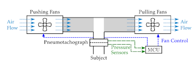
Schematic of fan-based FOT measurement device for free-breathing subjects


Results from a clinical trial on 60 subjects, that included healthy individuals as well as asthmatic and COPD patients, are also presented and, in the end, Dr. Maes offers a new framework for the time-varying behavior of the respiratory system during spontaneous breathing.

We congratulate Dr. Maes on this achievement and very interesting approach. This elegant work contributes towards the advances required to ultimately enable routine provision of the low-frequency FOT in patient populations and for clinicians to benefit from its diagnostic capacity.

For more information on the work of Dr. Maes and access to the full manuscript, please click here.


1. Hantos, Z., Daróczy, B., Suki, B., Galgóczy, G. & Csendes, T. Forced oscillatory impedance of the respiratory system at low frequencies. J. Appl. Physiol. 60, 123–32 (1986).
2. Hall, G. L., Hantos, Z., Wildhaber, J. H. & Sly, P. D. Contribution of nasal pathways to low frequency respiratory impedance in infants. Thorax 57, 396–9 (2002).
3. Maes, H. Low Frequency Forced Oscillation Technique in Clinical Practice. (Vrije Universiteit Brussel, Belgium, 2017).


Monday, March 5, 2018

SCIREQ INTRODUCES DRY POWDER DELIVERY AT SOT / TOXEXPO 2018

Many novel inhaled compounds and drugs require dry powder delivery and preclinical testing to assess efficacy and safety. The inExpose is an inhalation exposure system that provides a small-scale, configurable exposure for mice and rats studies.

Our recent addition of dry powder delivery expands the range of exposure environments beyond cigarette smoke, electronic cigarette vape, aerosols and gases.


The dry powder integration with the inExpose features:
          »   Up to 4 towers or chamber to be exposed simultaneously
          »   Identical or varying levels of exposure per tower or chamber
          »   Existing automation and control
          »   Small- scale exposure, minimizing compound use


Will you be attending the Society of Toxicology meeting in San Antonio next week? If so, I would like to invite you to come by booth #1545 to see our solutions for your preclinical respiratory research. We will be presenting a range of precise preclinical physiology solutions for safety pharmacology and toxicology studies, including:

          »   Automated, small-scale inhalation exposure studies featuring
               cigarettes, e-cigarettes, dry powders and aerosols
          »   Non-invasive tracking of ventilatory patterns and controlled gas exposure 
               with whole body plethysmography
          »   Continuous monitoring of physiological parameters with implantable and
               jacketed telemetry

Exhibit hours - Booth 1545
          Monday March 12 9:15am to 4:30pm
          Tuesday March 13 9:15am to 4:30pm
          Wednesday March 14         9:15am to 4:30pm

Click here to set-up a meeting to learn more about our solutions for your safety pharmacology and toxicology studies. 

If you are not planning to attend the conference but would like to receive additional information about our respiratory equipment, please contact our team of application specialists.

Looking forward to seeing you in San Antonio!

Monday, February 26, 2018

flexiVent User Group Meeting - April 26, 218 in San Diego

Join us for a day of scientific presentations and discussions to learn more about the flexiVent’s capabilities & applications and share your feedback with other users. 


WHEN?        Thursday April 26, 2018
                       Following the Experimental Biology Conference 2018

WHERE?      San Diego, California (Hilton San Diego Bayfront)

AGENDA:      » Presentation & Demo by our flexiVent Experts
                       » Case Studies & Interactive Session
                       » User Presentation: Learn how others use the flexiVent
                       » Recent Developments and a chance to ask your questions

Final Agenda and Details coming soon!  Space is limited, please CLICK HERE to register and be part of this event.

If you are interested in presenting your pulmonary research work during this user group, do not hesitate to contact us as soon as possible, to include your presentation in the agenda.

Please note that this user group meeting will be free of charge.

If you have any questions, do not hesitate to contact us at [email protected] or toll-free at 1-877-572-4737.

We look forward to seeing you in San Diego!

Best regards,
Your SCIREQ Team

Wednesday, February 21, 2018

SCIREQ at AAAAI & WAO Joint Congress in Orlando

Will we see you at the American Academy of Allergy Asthma & Immunology (AAAAI) conference in Orlando, next week? This congress is a wonderful opportunity to convene with other world-class researchers to discuss the latest discoveries in asthma and inflammatory diseases.

Mouse models have been and continue to be of invaluable importance in asthma research, for the elucidation of key disease pathways as well as in the assessment of potential therapies as highlighted in recent articles.

SCIREQ enables scientists to measure detailed lung function outcomes utilizing the flexiVent, which has become the gold-standard for in vivo respiratory research.

Join us at booth 229 to learn more.                                   

Exhibit Hours
Saturday, March 3:   9:45am – 3:15pm
Sunday, March 4:     9:45am – 3:15pm
Monday, March 5:  9:45am – 2pm


»      Click here to set-up a meeting with our specialists during or after the symposium.
»      Click here to view the AAAAI & WAO Joint Congress details.

If you are not planning to attend the conference but would like to receive additional information about our respiratory equipment, please contact our team of application specialists.


Monday, February 19, 2018

Upgrade to flexiWare 8.0

We are happy to announce the release of flexiWare 8.0!  This latest version is unified software for both the flexiVent and inExpose systems and flexiWare 8.0 includes features such as:


FLEXIVENT

  • Official release of the Lung Volumes maneuver along with its associated tasks and outcomes!
  • Diagnostic & power-on Self-Test available, allowing for greater confidence in the system and easier troubleshooting.
  • Significantly reduced time to load and view data in review mode (retroactively available for previously collected data as well).
  • Pop-up now appears on screen to notify users of excluded datasets as they occur to allow for immediate action.
  • Ability to copy subjects when creating new subjects or studies.

INEXPOSE

  • System leak test available with easy to follow software wizard to ensure the system is leak free prior to beginning an experimentation session.
  • Ability to reinitialize and modify CSR setting anytime during an experimentation session.
  • Significantly improved and integrated profile editor, including the ability to view pump and nebulizer trigger signals in real-time.
  • Scripting capabilities added for complex protocols as well as for improved standardization.
  • Ability to create export scenarios for easier data export.

Ready to upgrade?

Contact us to find out if you are eligible for a free upgrade and how to have flexiWare 8.0 running in your lab!

Phone: 1.514.286.1429
Toll Free: 1.877.572.4737
Email: [email protected]

Monday, January 22, 2018

Refined Study Design with Spirometry

Spirometry is a widely known clinical pulmonary test measuring volumes and flows expired by patients that is used to confirm a diagnostic of respiratory disease or follow treatment. SCIREQ’s flexiVent’s offers an analogous test for preclinical research with its Negative Pressure Forced Expiration (NPFE) extension. By exposing the subject’s airway opening to a negative pressure to rapidly draw out air from the lungs, the system can generate flow-volume loops or volume-time plots, from which the flexiWare software can then extract clinically relevant volume and flow parameters (e.g. FEVx, FEFx, FVC, PEF).

A recent publication by Devos et al.1 characterized forced expiration measurements in some well-established mouse models of lung diseases, which specific phenotypes were confirmed by a concomitant respiratory mechanics assessment. The researchers observed that disease-induced changes in forced expiration-related charts and parameters were generally similar to what was observed in the clinic. For example, when compared to a control group of healthy mice.

  • Mice with fibrosis exhibited a typical restrictive profile, with a reduced PEF & FVC and a normal FEV0.1/FVC ratio. 
  • Mice with emphysema displayed a decrease in PEF characteristic of an obstructive phenotype.
  • Mice presenting an acute lung injury had significantly reduced PEF.
  • Mice with features of asthma showed a decrease in FEV0.1 following methacholine challenges.

SCIREQ’s flexiVent is the first platform that allows forced expiratory manoeuvres to be performed side-by-side with respiratory mechanics, pressure-volume loops, or lung volume measurements in mice and rats. This unique feature enables a refined study design with outcomes susceptible to translate between preclinical research and clinical settings. 


Read more
1Devos, FC et al. 2017. Forced expiration measurements in mouse models of obstructive and restrictive lung diseases. Respiratory Research 18: 123.

http://blog.scireq.com/2017/11/comprehensive-lung-function-assessments_27.html

Leaving Nothing Out

The functional residual capacity (FRC) plays an important role in maintaining optimal ventilation.  FRC is a subdivision of the total lung capacity (TLC) and represents the volume of air left in the lungs at the end of an expiration.  It is involved in keeping the small airways open for an efficient gas transfer between the air and the blood.  Variations in that volume can be seen under several physiological or pathophysiological conditions and can be associated with clinical symptoms like dyspnea. 

FRC is a clinically important parameter which is determined, along with other lung volumes/capacities, when performing a full lung function assessment.  It is inversely related to airway resistance, such that a reduction in FRC will increase the level of opposition to airflow.  At the preclinical level, while measurements of airway resistance are routinely performed in many research laboratories, the determination of FRC is rarely reported as it can prove to be technically challenging, especially in subjects as small as mice.  

It is possible to obtain FRC from a full-range pressure-volume (PV) curve.  This was done until now in mice by determining the volume at a conventionally or arbitrarily defined equilibrium pressure (e.g. 0 cmH20), since FRC represents the volume at rest.  In a recent report1, FRC was estimated by a novel approach in rodents, following the automation of the full-range PV curve technique with SCIREQ’s flexiVent system.  It was estimated by volume subtraction and took advantage of the access to varied, precisely controlled, automated maneuvers within a single device.  In comparison to the previously established methodology, this novel approach demonstrated a more accurate estimation of FRC, particularly in diseased subjects.

The estimation of FRC by volume subtraction can be performed in rats and mice along with the determination of other lung volumes/capacities, measurements of respiratory mechanics, or partial PV curves.  The authors showed that the FRC estimate more than tripled in a mouse model of emphysema, representing the most important change in a panel of lung volumes/capacities.  Including measurements of FRC and other lung volumes/capacities in the characterization of respiratory models or novel therapeutic strategies can contribute to generate comprehensive and robust functional assessments. 

No model fully recapitulates human respiratory diseases.  Let’s reconsider the endpoints typically reported so that nothing that matters gets ignored.


Read more
1Robichaud, A et al. 2017. Automated full-range pressure-volume curves in mice and rats. Journal of Applied Physiology 123: 746-756.

Monday, January 8, 2018

Empower yourself

Attending a training such as the Phenotyping Mice Models of Human Lung Disease organized by the Jackson Laboratory is a great way to advance skillsets or simply get started in the field of respiratory research. It represents a wonderful opportunity for researchers of all stages to learn and network in a structured yet informal environment. The uniqueness of the event lies in the roundness of the approach, which combines theoretical sessions and practical experience in all topics.

SCIREQ has been a partner in this event for years now and has contributed with providing hands-on experience to participants with lung function measurements using the flexiVent alongside authorities in the field. Practical exercises are designed to demonstrate a response to a particular intervention, highlight unique measurements (including partitioning the lungs between airway and tissue effects), and analyze outcomes with respect to their physiological implications.

JAX Experiments
As an example, participants were guided during the last workshop to utilize pressure-volume loops to confirm the effects of an intervention with implications on lung surfactant and atelectasis.  
Figure 1: Pressure-volume loops from a subject before (left) and after (right) a lung lavage. The red line represents the Salazar-Knowles equation fit to the deflation limb of the pressure-volume curve.
They were also invited to examine the effect of increasing the positive end expiratory pressure (PEEP) during ventilation and lung function measurements while exploring the impact on detailed respiratory mechanics parameters before and after the intervention.

Airway responsiveness to a specific bronchoconstrictor agent before and after a therapeutic treatment or an assessment of various lung volumes are also typically part of the repertoire of techniques taught.  


Figure 2: Single (left) and broadband (right) forced oscillation outcomes prior to and following increasing doses of nebulised methacholine in presence or absence of a bronchodilator treatment.

Looking forward
The event runs every second year in Bar Harbor, Maine, and accepts a limited number of participants. Plan ahead to take part in the workshop, which we highly recommend to both senior researchers and students alike.